Fig. 1.
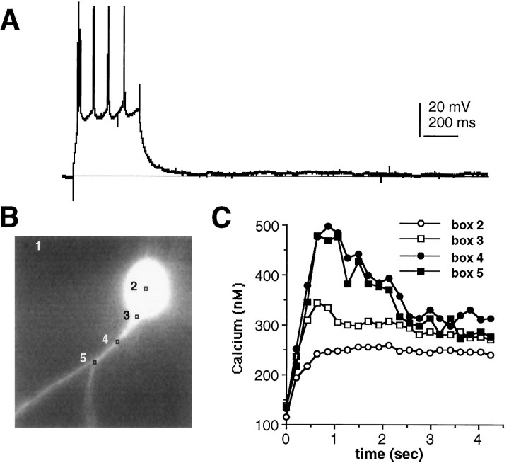
Calcium homeostasis of DLSN neurons.A, Spike train elicited by a depolarizing current pulse (0.2 nA, 400 msec). Membrane potential was held at −80 mV.B, Fluorescence image of this DLSN neuron, 380 nm excitation. Analysis locations are indicated by 3 × 3 pixel square boxes. Each pixel subtends an area of ∼0.5 × 0.5 μm. Box 1 is used as background to correct the 350/380 ratio measured atbox 2–5. C, Calcium increases in soma and dendrites caused by firing are shown in A.

