
Fig. 7.
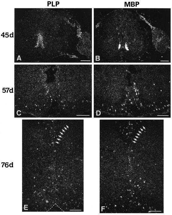
Origin and dispersion of cells expressing PLP/DM20 and MBP transcripts in the spinal cord. Expression pattern of PLP/DM20 (labeled PLP in A, C, E) and MBP (B, D, F) transcripts at 45, 57, and 76 dpc of human spinal cord development, as revealed by in situ hybridization using 35S-labeled riboprobes on cryostat sections. Two foci of PLP/DM20 or MBP transcripts are detected in the ventral cord at 45 dpc (A, B). Note that the domain of expression of PLP/DM20 transcripts at this age is wider and spreading more dorsally into the VZ, whereas the MBP transcripts seem to coincide mostly with the two clusters of oligodendrocyte precursors detected with MBP and O4 antibody staining (see Figs. 1B, 2A). Note a discrete but definite signal for these transcripts in the dorsal (B) and ventral roots (A) on theright of the cord sections. At 57 dpc, clusters of PLP/DM20 and MBP transcripts, probably associated with cells, are now dispersed in the ventral forming white matter (C, D), and at 76 dpc (E, D) they become detectable in both the dorsal (on the left of the row ofshort arrows in E and D) and ventral forming white matter. (Such transcript clusters could also be detected in the lateral spinal cord at this age; not illustrated here.) Scale bars, 200 μm.