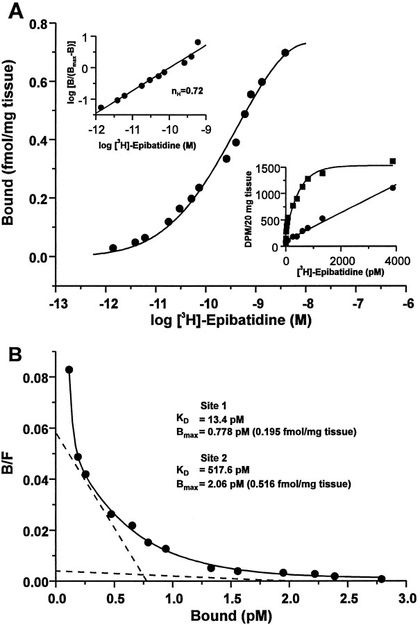
Fig. 1.
Neuronal nicotinic receptor binding sites in the rat trigeminal ganglion. Saturation binding analysis of neuronal nicotinic receptors labeled with [3H]-epibatidine in membrane homogenates of rat trigeminal ganglion. Aliquots of homogenized, washed trigeminal ganglion membranes equivalent to 20 mg of original tissue weight were incubated in triplicate with [3H]-epibatidine at the indicated concentrations (1.4 pm to 3.9 nm) in the absence or presence of 300 μm nicotine bitartrate (to define nonspecific binding). The median value of each triplicate measurement was used to generate all data. A, Semilog plot of the data expressed as bound receptor (fmol/mg tissue) and calculated as the difference between total and nonspecific binding. Top left inset, Hill plot, including the calculated Hill coefficient (nH), of the transformed data. Bottom right inset, Linear plot of the specific (squares) and nonspecific (circles) binding at each concentration of [3H]-epibatidine.B, Rosenthal plot of the data shown in A. Data were analyzed by nonlinear regression with LIGAND (Munson and Rodbard, 1980) and were best fit to a two-site model (ANOVA,F1,8 = 51.9; p < 0.001), as indicated by the dashed lines. The calculated receptor density (Bmax) and equilibrium dissociation constant (KD) values for each site are indicated. Data are representative of an experiment performed four times.

