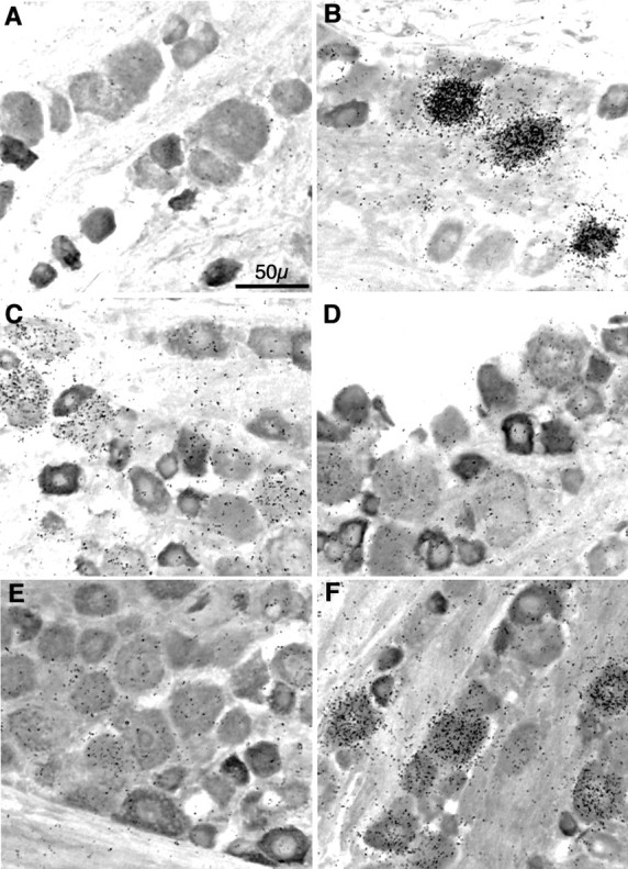
Fig. 2.

Neuronal nicotinic receptor subunit mRNA expression in sensory neurons of the rat trigeminal ganglion. Combinedin situ hybridization histochemistry and immunocytochemistry for neuronal nicotinic receptor subunit mRNAs and peripherin in the rat trigeminal ganglion. Bright-field images are of 20-μm-thick frozen sections of adult male rat trigeminal ganglion sequentially processed for in situ hybridization by a35S-labeled riboprobe complimentary to the mRNA encoding the α2 (A), α3 (B), α4 (C), α5 (D), β2 (E), or β4 (F) neuronal nicotinic receptor subunit (represented by black grains), followed by immunocytochemistry with an anti-peripherin rabbit serum (seen asdarkly stained cells). After ABC (peroxidase) color development, slides were subjected to emulsion autoradiography, counterstained, coverslipped, and photographed. Total magnification, 40×; scale is indicated in A. Photographs are representative of experiments performed at least five times for each subunit transcript with two separate and nonoverlapping probes.
