
Fig. 5.
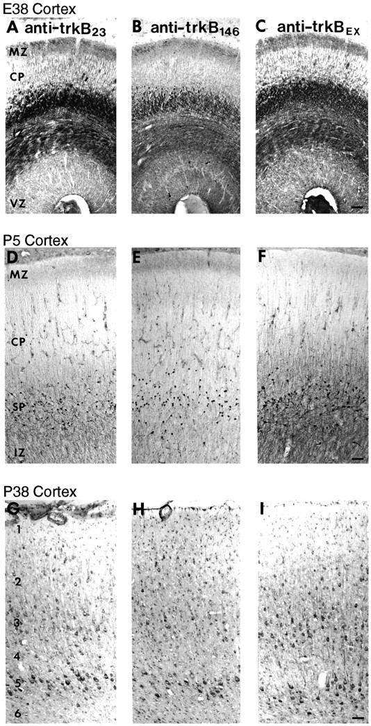
Comparison of cortical distribution of trkB-immunoreactive cells and fibers using three distinct anti-trkB antibodies over a wide range of developmental ages.A–C, Coronal sections from an E38 ferret stained with 0.5 μg/ml anti-trkB23 (A), 1 μg/ml anti-trkB146 (B), or 2.8 μg/ml anti-trkBex (C). D–F, Sagittal sections from a P5 ferret stained with 0.5 μg/ml anti-trkB23 (D), 2 μg/ml anti-trkB146 (E), or 2.8 μg/ml anti-trkBex (F). G–I, Sagittal sections from a P38 ferret stained with 1 μg/ml anti-trkB23 (G), 2 μg/ml anti-trkB146 (H), or 1.4 μg/ml anti-trkBex (I). CP, Cortical plate; IZ, intermediate zone;MZ, marginal zone; SP, subplate;VZ, ventricular zone; 1, cortical layer 1; 2, layer 2; 3, layer 3;4, layer 4; 5, layer 5; 6, layer 6. Scale bars: A–C, 55 μm; D–F, 55 μm; G–I, 55 μm.