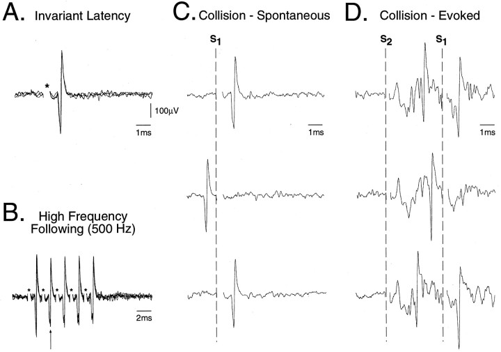
Fig. 1.
Criteria used to antidromically identify TGT neurons. Five superimposed oscilloscope traces are presented of an antidromic action potential recorded in the rostral trigeminal sensory nuclear complex (TSNC) after stimulation of the thalamus using (A) single pulse (95 μA, 0.1 msec, 1 Hz) and (B) a high-frequency (500 Hz, 5 pulses) train of stimuli. The asterisks in A andB denote the stimulus onset. Consecutive antidromic responses displayed constant latency-to-onset indicating that the action potential resulted from antidromic activation of the axon of the recorded unit. Note that during the high-frequency train inB, a progressive increase in the soma–dendritic conduction time is apparent (arrow), indicating that successive antidromic spikes were generated within the relative refractory period for this cell. Both spontaneous (C) and evoked (D) types of collision are presented. InC, single oscilloscope sweeps illustrate a collision between the antidromic spike and a spontaneous action potential. The sweeps are aligned by the onset of thalamic stimuli (S1) as indicated by the dashed vertical line. In D, single oscilloscope sweeps illustrate a collision between an orthodromically activated action potential evoked by stimulation of the IAN (S2: 0.2 msec, 150 μA) and the antidromic spike. The extracellular field potential evoked by IAN stimulation in D indicates that this neuron was located within the boundaries of the TSNC (Cairns et al., 1995).

