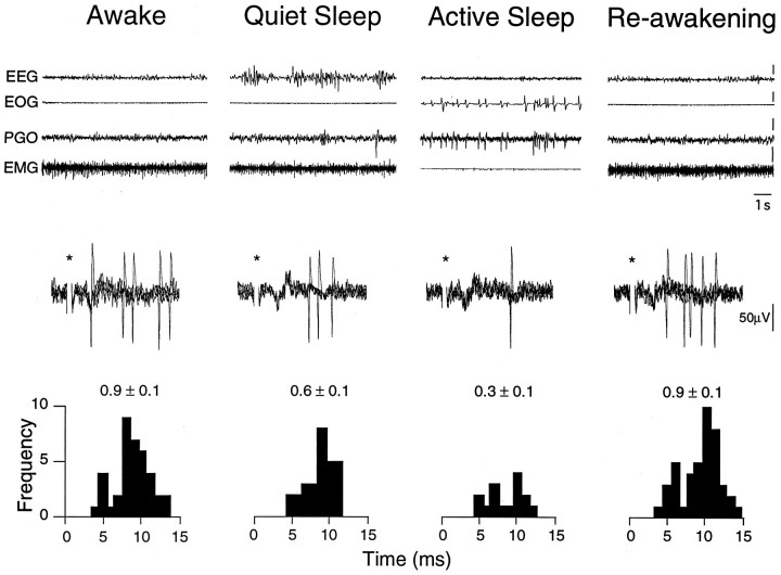
Fig. 6.
Active sleep-related suppression of tooth pulp-evoked TGT neuronal activity. Oscilloscope traces represent behavioral state and neuronal activity as described in Figure 5. PSTHs were constructed from 50 consecutive responses to low-intensity bipolar electrical stimuli applied to the canine tooth pulps (0.2 msec, 12 μA, 1 Hz). The number above each PSTH indicates the mean evoked activity (in spikes per stimulus ± SE). Note that tooth pulp-evoked activity in this neuron was reversibly suppressed by 33% during QS and 67% during AS when compared with W.

