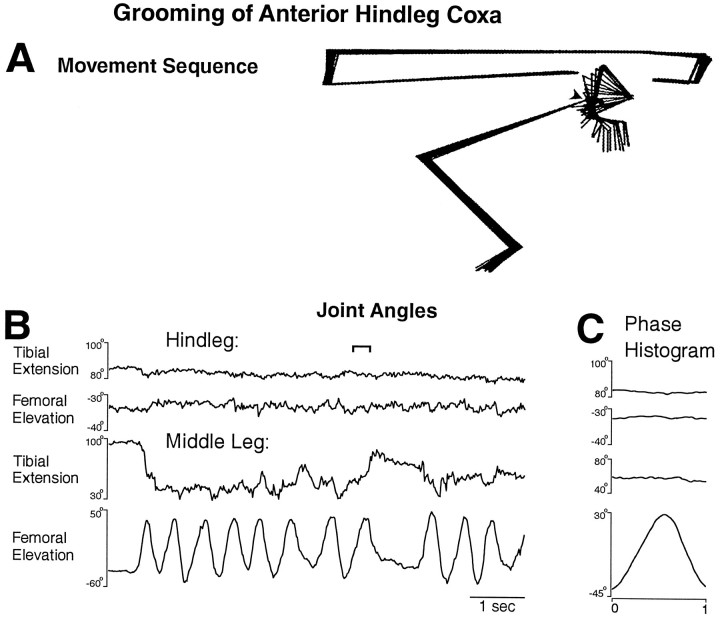
Fig. 4.
Middle leg grooming of the anterior hindleg coxa.A, Stick figure sequence for the time period indicated by the bracket in B. B, Hindleg and middle leg tibial and femoral joint angles as a function of time during an episode of grooming of the anterior hindleg coxa.C, Phase histogram of joint angles for the episode of grooming shown in B, generated from 12 cycles. The reference joint angle was middle leg femoral elevation. This is the same animal as shown in Figures 1, 2, 3.

