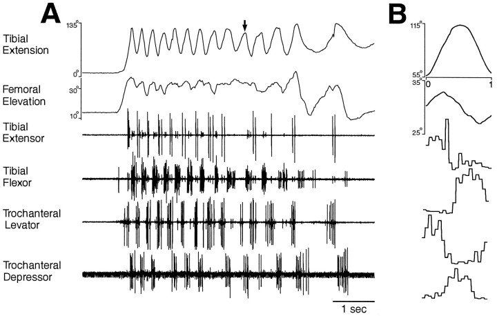
Fig. 6.
Hindleg joint angles and simultaneous hindleg EMG recordings for hindleg grooming of the posterior abdomen. Fromtop to bottom, traces are tibial extension angle, femoral elevation angle, tibial extensor EMG, tibial flexor EMG, trochanteral levator EMG, and trochanteral depressor EMG.A, Joint angles and EMGs as a function of time.Arrow indicates a cycle of tibial extension that occurred without recorded tibial extensor muscle activity.B, Phase histogram of joint angles and EMGs for the episode of grooming shown in A, generated from 13 cycles. The reference joint angle was tibial extension.

