
Fig. 9.
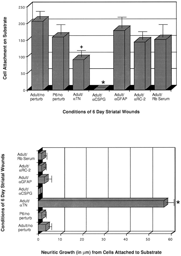
Histograms showing TH+ cell attachment (top graph) and process outgrowth (bottom graph) on >5 d wounds treated with antibodies or serum. Note the dramatic reduction in TH+ cell attachment on both CS-56 and anti-tenascin-treated cultures, as compared with untreated adult cocultures (Adult/no perturb), anti-GFAP, anti-RC-2, and rabbit serum-exposed explants that exhibit no statistically significant differences from the adult or postnatal day 6 untreated cultures (n = 8 for the adult/no perturb;n = 3 for anti-GFAP; n = 6 for the anti-tenascin experiments). Reduced cell attachment on anti-tenascin-treated cocultures was accompanied by an increase in neurite outgrowth. Wound substrates from the P6 striatum maintain a similar level of cell attachment and process outgrowth of E15 VM cells, as do their adult counterparts. (+) and (*) denote statistically significant differences from adult/nonperturbated (nontreated, control) conditions, with p > 0.025 andp > 0.005, respectively.