
Fig. 6.
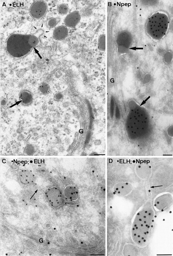
Electron micrographs of a plastic section (A) and ultrathin cryosections (B–D) of the TGN of type I neurons. Within condensing vacuoles at the trans-Golgi (B) and at various sites in the TGN (A), condensed protein cores with distinct electron densities are found (bold arrows). The lighter part labels for ELH (A), and the darker portion labels for N-terminal peptide (B). The membranes surrounding these differentially condensed proteins may form coated buds (small arrows in A). Sometimes protein cores with similar (C) or distinct (D) protein contents were segregated within the continuous membrane of the TGN (arrows). Scale bars, 0.1 μm.