
Fig. 1.
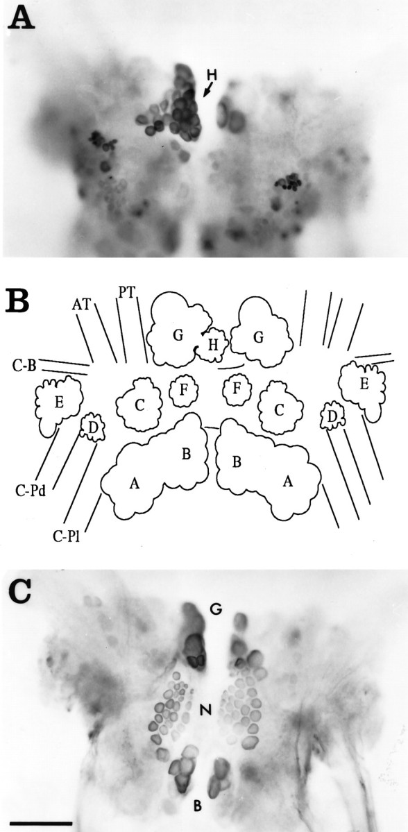
Whole mounts showing the locations of neurons containing CP2-lir in the cerebral ganglia visualized using GAR-biotin and ABC-HRP as viewed from the ventral surface.A, Dorsal surface. The location of the Hcluster is indicated (arrow). B, Diagram of the dorsal surface illustrating the positions of the major neuronal clusters. C, Ventral surface. The labels forG, N, and B clusters are positioned between these bilateral clusters. Immunoreactive neuronal cell bodies are located in a number of neuronal clusters on both sides of the ganglia (see text). Photomicrographs and diagram are as viewed from the ventral side, so right and left ganglia appear reversed forA and B. Neuronal clusters are designated as in Jahan-Parwar and Fredman (1976), except the Ncluster (see text). AT, Anterior tentacular nerve;C-B, cerebral–buccal connective; C-Pd, cerebral–pedal connective; C-Pl, cerebral–pleural connective; PT, posterior tentacular nerve. Scale bar (shown in C): 200 μm (applies toA–C).