Fig. 10.
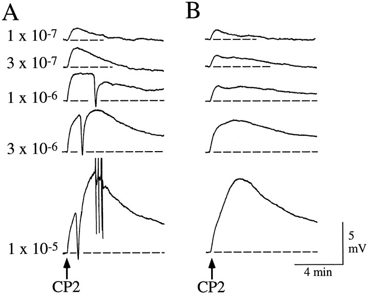
Responses of neurons in the abdominal ganglion to bolus applications of CP2. A, Depolarization of R20 by bolus application of CP2 at the indicated concentrations. R20 was identified by relative size and position, pigmentation, pronounced spike broadening exhibited during repetitive firing, synaptic connections to RB or RG neurons, and the nature of its responses to applications of FMRFamide and myomodulin (Alevizos et al., 1989). This experiment was performed in nASW. Note that, in addition to a dose-dependent depolarization of R20, the higher doses of CP2 also recruited a large hyperpolarizing response that was made up of a number of individual IPSPs when observed at a faster time base.B, Depolarization of R20 by bolus application of CP2 at the indicated concentrations. This experiment was performed in low Ca ASW to suppress chemical synaptic transmission. CP2 continued to cause a dose-dependent depolarization similar in amplitude and time course to those observed in A, but no compound IPSPs were recruited. Records in A and B are from the same neuron. Similar results were obtained from R20 neurons in other preparations (11 of 11 analyzed).

