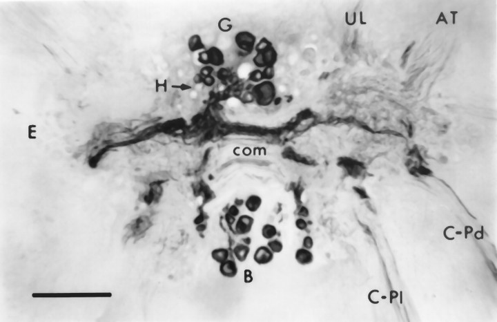
Fig. 2.
Section through the cerebral ganglia near ventral surface showing CP2-lir in the cytoplasm of neuronal cell bodies in the regions of the B, G, and Hclusters visualized using GAR-biotin and ABC-HRP. Large immunoreactive fibers extend from these clusters and extend across the commissure (com) and leave the ganglion in the cerebral–pedal (C-Pd) and cerebral–pleural (C-Pl) connectives. Smaller fibers with varicosities are found throughout the neuropil. AT, Anterior tentacular nerve; E, E cluster;UL, upper lip nerve. Photomicrograph is viewed from the ventral side, so right and left ganglia appear reversed. Scale bar, 200 μm.

