
Fig. 5.
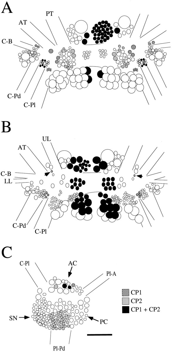
Schematic diagrams of the location of neuronal cell bodies containing CP1-lir or CP2-lir, or both CP1-lir and CP2-lir in selected ganglia. A, Dorsal surface of the cerebral ganglia. B, Ventral surface of the cerebral ganglia.Arrows indicate the positions of isolated individual neurons in the M clusters that contained CP1-lir only.C, Medial surface of the right pleural ganglion. Nerves from the cerebral ganglia are designated as in Fredman and Jahan-Parwar (1976). AC, Anterior cluster; AT, anterior tentacular nerve; C-B, cerebral–buccal connective; C-Pd, cerebral–pedal connective;C-Pl, cerebral–pleural connective; LL, lower lip nerve; PC, posterior cluster;Pl-A, pleural–abdominal connective;Pl-Pd, pleural–pedal connective; PT, posterior tentacular nerve; SN, sensory neuron cluster;UL, upper lip nerve. Scale bar, 200 μm (applies toA–C).