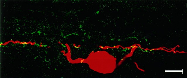
Fig. 15.
Top. A double-labeled vertical section containing a Neurobiotin-injected ON-parasol ganglion cell (red) and amacrine cell processes labeled with antisera to ChAT (green). The inner set of cholinergic processes was narrowly costratified with the ON-parasol cell dendrites. This confocal image represents a reconstructed stack of optical sections spanning 1 μm in depth. Scale bar, 25 μm.

