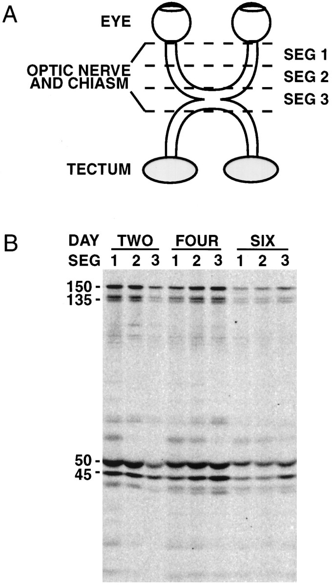
Fig. 2.

Association of dynactin with axonal transport slow component b. Segmental analysis was performed to confirm the association of dynactin with SCb. Axonally transported proteins were radiolabeled with Tran35S-label via intravitreal injection.A, The radiolabeled optic nerves and tracts were removed at specified times after injection and divided into three ∼5 mm segments, as diagramed. Segment 1 corresponds to the proximal half of the optic nerve. Segment 2 corresponds to the distal half of the optic nerve. Segment 3 corresponds to the optic chiasm and the proximal portion of the optic tract. The segments were homogenized in lysis buffer. Dynactin was then immunoprecipitated and analyzed by SDS-PAGE and storage phosphor autoradiography. B, An autoradiograph showing the radiolabeled dynactin polypeptides immunoprecipitated from segments of the optic nerve and tract at the indicated times after injection. Above each set of three lanes is the number of days (DAY) after injection that the optic nerve and tract were isolated (TWO,FOUR, and SIX). Numerals (1, 2, 3) indicate the isolated segment (SEG) of the optic nerve or tract analyzed in that lane. The major dynactin subunits and isoforms are indicated to the left of the gel: p150Glued (150), p135Glued (135), p50 (50), and Arp1 (45). Increasing amounts of each of the dynactin subunits are seen in the more distal segments with increasing time after injection.
