Fig. 2.
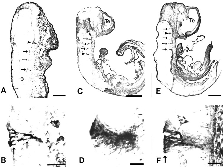
Tropomyosin is expressed at rhombomere boundaries in the developing rat hindbrain. Parasagittal sections at E10 (A, B), E11 (C,D), and E12 (E, F) labeled with the anti-tropomyosin TM311 illustrate several TM311-immunoreactive boundaries between rhombomeres in the hindbrain (filled arrows andarrowheads in A, C,E; boundaries shown at higher magnification inB, D, F are indicated byarrowheads). At E10, corresponding to the onset of rhombomere formation, the boundaries are narrow and lightly immunoreactive compared to later ages. Open arrow inA indicates a recently formed boundary that shows TM immunoreactivity in adjacent sections. Higher-magnification photomicrographs of individual rhombomere boundaries (B,D, F) show that TM311-immunoreactive boundaries are 4–8 cells wide and that the cells extend across the width of the neuroepithelium. Arrowsin B and F indicate locations of cell bodies. In the CNS, TM311 immunoreactivity is also evident in the smooth muscle of blood vessels and in connective tissue (F). Scale bars: A, 145 μm;B, D, E, 35 μm;C, E, 625 μm.

