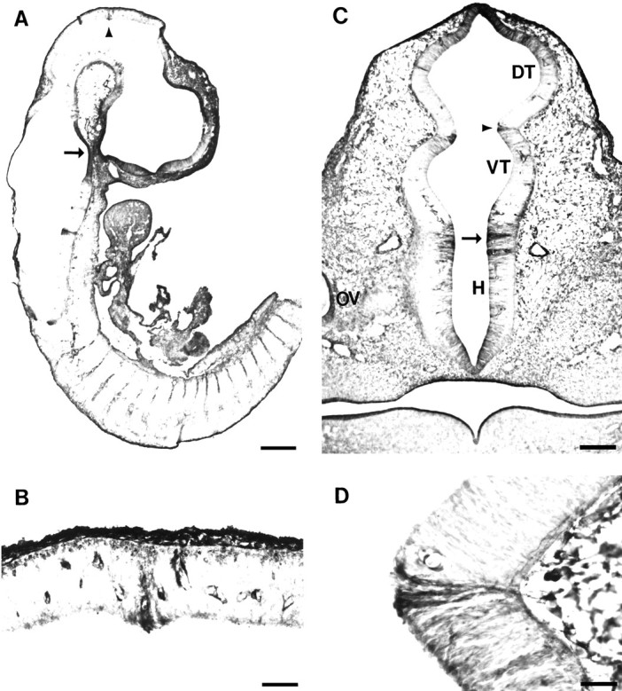
Fig. 3.

TM311 labels some prosomere boundaries.A, Midsagittal section through E12 rat showing TM311 immunoreactivity localized to the pretectum/tectum boundary (arrowhead). Note the TM immunoreactivity in dorsal root ganglia and intersomitic mesenchyme. B, Higher magnification of pretectum/tectum boundary (arrowhead in A), with TM311 immunolocalization that distinguishes the caudal boundary of prosomere P1. Note the blood vessel staining on either side of the TM311-positive boundary. C, Coronal section through E12 rat diencephalon at the level of the optic vesicle (OV) showing TM311 immunolocalization at the boundary between the dorsal thalamus (DT) and ventral thalamus (VT) (arrowhead, P2–P3 boundary). A boundary is also suggested between the VT and hypothalamus (H; arrow), contrasting the P3–P4 prosomeres. D, Increased magnification of TM311 immunolocalization at the zona limitans intrathalamica, the border between the DT and VT, a representation of the P2–P3 prosomeric boundary (arrowhead in C). Note the floor plate labeling in presumptive pons (arrow inA). Scale bars: A, 470 μm;B, D, 50 μm; C, 150 μm.
