Fig. 5.

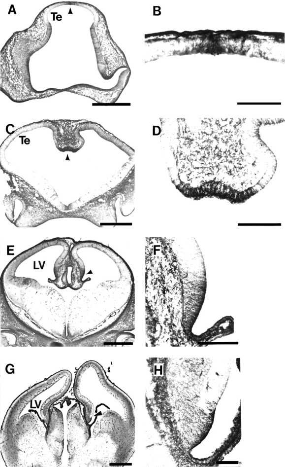
Tropomyosin immunolabels cells that are the forerunners of the choroid plexus. A, Coronal section through E11 rat showing TM311-immunolabeled cells (arrowhead) at the dorsal midline of the telencephalon (Te) that will form the choroid plexus 2–3 d later.B, Higher magnification of the restricted zone of TM311-labeled neuroepithelium (arrowhead inA). C, Coronal section through E12 rat telencephalon showing intensely TM311-immunoreactive cells (arrowhead) at the ventral-most region of the medial wall of the dorsal telencephalon. These cells will form the choroid plexus of the lateral ventricles. D, Higher magnification of the TM311-immunoreactive choroid plexus precursors (arrowhead in C). E, Coronal section through E14 rat showing TM311-labeled choroid plexus (arrowhead) of the lateral ventricle (LV). F, Higher magnification showing TM311-labeled cells of the presumptive choroid plexus (arrowhead in E) distinguished from the TM311-negative presumptive allocortex. Note that the plexus has already formed in the LV and is TM311-positive (lower right side). G, Coronal section through the E16 rat telencephalon showing TM311-immunoreactive choroid plexus and choroid plexus precursors (arrowhead). H, Increased magnification showing TM311-positive cells of the developing choroid plexus at the ventro-medial wall of the dorsal telencephalon. Scale bars: A, C, E,G, 500 μm; B, 100 μm;D, F, H, 200 μm.
