
Fig. 6.
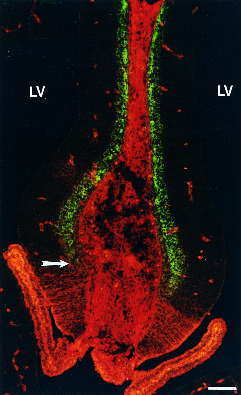
Color photomicrograph highlighting the molecular distinction between neuroepithelium-containing choroid plexus and allocortical precursors. Coronal section through E15 rat telencephalon reveals the sharp boundary (arrow) between the region containing the TM311-labeled presumptive choroid plexus (ventrally, inred) and allocortex, shown by MAP2-positive neurons in the primordial plexiform layer (dorsally, in green). TM is also expressed in the smooth muscle of the blood vessels and in connective tissue. Scale bar, 40 μm.