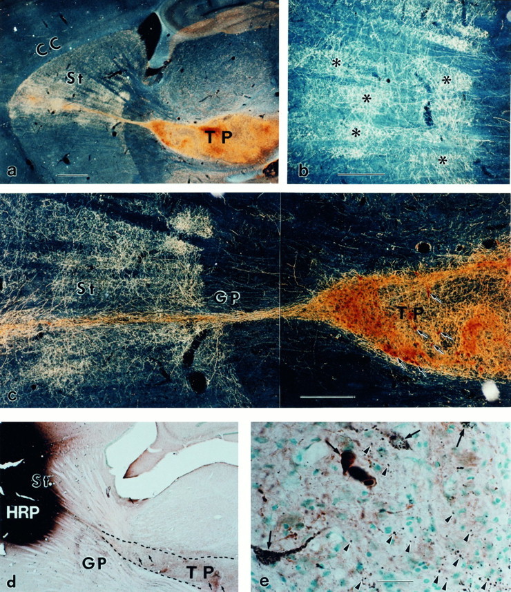
Fig. 1.

Striatonigral and nigrostriatal reconnections by the KA-bridged mesencephalic transplant. a, The TH-positive neurons in the bridged mesencephalic transplant (TP) sent bundled fibers along the KA track through the globus pallidus (GP). These fibers from the bundle expand immediately on arrival in the striatum (St).b, On arrival, the TH-positive fibers form patches (asterisks) in the striatum, which is similar to the developmental pattern of DA fibers in the immature St at midgestation. The patchy innervation (b) is transient and spreads into homogeneous innervation (c) (Zhou and Chiang, 1995) over a 3–6 week period after transplantation. The TH immunocytochemistry (brown) and HRP (black) double staining indicate that HRP injection in the St and the adjacent cortex (d) results in retrograde transport labeling of the neuronal cell bodies in the TP; many of them are HRP- and TH-double-labeled (e, arrows) (in this small injection site, 1432 TH-positive neurons were observed in transplant; among them ∼13.7% were HRP-positive). The TH and HRP double labeling of the cell bodies in the TP indicate that DA neurons in the bridged TP sent fibers to the St and/or cortex. The anterograde transport labeling also results in punctate terminal staining in the TP (e,arrowheads). These punctate HRP granules are located outside the TH-positive fibers, indicating that they are not retrogradely transported labeling through DA fibers but rather antegradely transported through fibers of striatal neurons into their terminals within the TP. This suggests that the striatal neurons also sent fibers that terminate in the TP. a–c, Dark-field photographs; d, e, bright-field photographs. CC, Corpus callosum. Scale bars:a, d, 1 mm; b,c, 0.5 mm; e, 30 μm.
