Fig. 4.
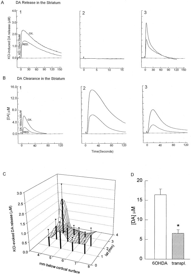
Transplantation of fetal mesencephalic tissue into the SN followed by KA bridging from the SN to the striatum restores the release and clearance of DA in the striatum 6–8 weeks after transplantation. A1, Local application of KCl (70 mm, 125 nl) (arrows) to the control nonlesioned striatum induces DA release. KCl is applied locally through a multibarrel pipette placed in the striatum of the rat. Extracellular DA concentration is recorded using Nafion-coated carbon fiber electrode. Upper trace, OX represents the extracellular signal from DA oxidation. The ratios of reduction (lower trace, RED) to oxidation currents are used to qualitatively identify the electroactive species as DA.A2, K+-induced DA overflow is abolished in the lesioned striatum and is restored in the lesioned striatum with a KA-bridged transplant (nigra to striatum) (A3). The peak of DA overflow (mean ± SEM) induced by K+stimulation is restored in the 6-OHDA-lesioned striatum from six transplanted animals. B1, Local application of DA (200 μm, 200 nl) (arrows) to the control nonlesioned striatum resulted in a retention of ∼6 μmDA overflow. B2, The extracellular DA concentration is increased after a 6-OHDA lesion (B2 vs B1) and is reduced in the lesioned striatum with a KA-bridged transplant (B3).C, A much broader area of DA release in the lesioned striatum is achieved by the KA-bridged transplant. A 2 × 3 × 3 mm striatal field is found responsible to K+ to release DA, with a higher release near the needle track.D, Restoration of DA clearance in transplanted animals.Open and filled bars represent peak extracellular DA concentrations in the lesioned and bridged striatum from six animals, respectively. The KA-bridged transplant diminishes the increase in DA overflow induced by local application of DA into the lesioned striatum (*p < 0.01, ttest).

