
Fig. 1.
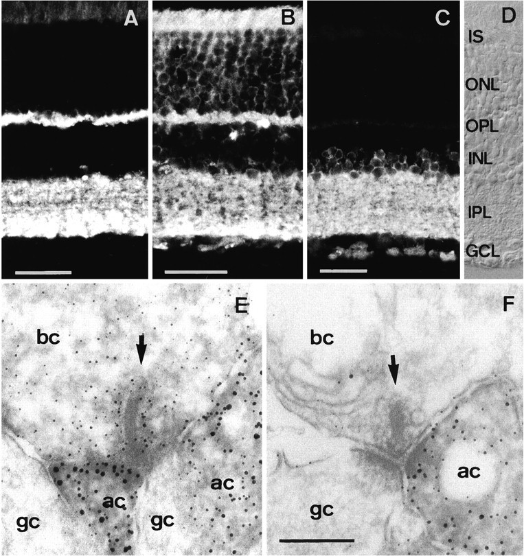
Vertical cryostat sections through rat retina labeled with antibodies against different synaptic proteins. Synaptobrevin (A) and SNAP-25 (B) are both present in the OPL and IPL. C, Syntaxin 1 is present only in the INL and IPL. D, Nomarski image showing the retinal layers. Electron microscopic localization of (E) SNAP-25 and (F) syntaxin 1 in the rat retina. Whereas SNAP-25 is present at all synapses of all neurons in the retina, syntaxin 1 is present only in amacrine cells that form conventional synapses. IS, Inner segments;ONL, outer nuclear layer; OPL, outer plexiform layer; INL, inner nuclear layer;IPL, inner plexiform layer; GCL, ganglion cell layer; bc, bipolar cell; ac, amacrine cell; gc, ganglion cell. Scale bars:A–C, 30 μm (scale bar inC also applies to D); E, F, 0.2 μm (shown in F).