Fig. 6.

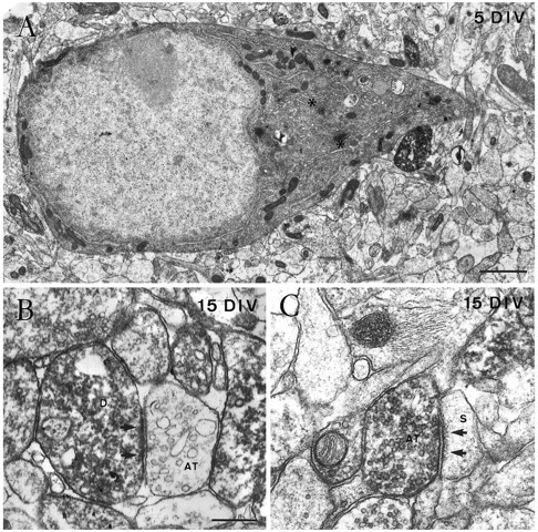
Fine structure of Cajal–Retzius cells in single hippocampal slice cultures, as shown by calretinin immunolabeling.A, Electron micrograph of the perikaryon of a Cajal–Retzius cell in the stratum lacunosum-moleculare after 5 DIV, showing a nucleus rich in euchromatin, a prominent nucleolus, and a cytoplasm rich in organelles such as the Golgi complex (asterisks). B, An immunopositive dendrite (D) close to the hippocampal fissure receives an asymmetric synaptic contact (arrows) from an unlabeled axon terminal (AT) after 15 DIV.C, Electron micrograph illustrating an immunoreactive axon terminal (AT) in the stratum lacunosum-moleculare in asymmetric synaptic contact (arrow) with an unlabeled dendritic spine (S) at 15 DIV. Scale bars: A, 1 μm;B, 0.4 μm, also pertains to C.
