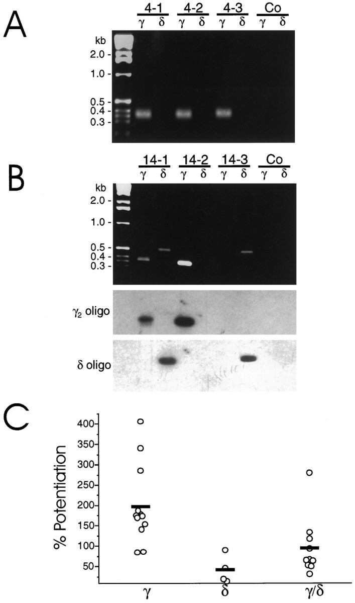
Fig. 5.

Single-cell RT-PCR analysis in cultured cerebellar granule cells. Granule cells from 4 DIV (A) or 14 DIV (B) cultures were analyzed by single-cell RT-PCR as described in Materials and Methods. A andB show three representative cells from each culture. For each PCR experiment, controls were performed to verify the absence of contaminating cDNA. Shown here are controls (Co) in which whole-cell recording was not performed, but a small amount of medium was removed from the bath solution with the patch pipette.B, Bottom panels show Southern blot analyses of the corresponding PCR products using radiolabeled γ2-specific primer 5′-AGCAACCGGAAACCAAGCAAGGATAAAGAC, which was then stripped and the membrane was rehybridized with the δ-specific primer 5′-TCAATGCTGACTACAGGAAGAAACGGAAAG. C shows the potentiation of GABA-activated currents induced by 1 μmTHDOC for individual cells at 14 DIV in which RT-PCR of the individual cells indicated the presence of γ and/or δ subunit mRNA as indicated. Mean potentiations of cells in which the presence of δ and γ/δ subunit mRNA were detected were statistically lower (p < 0.01) than that for cells in which only the γ subunit mRNA was found.
