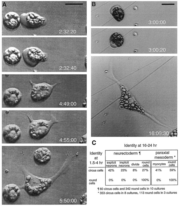
Fig. 3.

Morphologically differentiated neurons and myocytes are derived from persistent circus cells in culture.A, Circus cell from the neurectoderm that differentiates morphologically as a neuron. The neurectoderm cell at leftlacks membrane excursions and fails to differentiate morphologically.B, Circus cell from the paraxial mesoderm that differentiates as a myocyte. Times (after culturing) of image acquisition are noted at lower right of each frame. Scale bars, 25 μm. C, Differentiated fates of persistent circus cells from neurectoderm and paraxial mesoderm cultures are indicated as the percentage of the initial circus and round cell populations. Video time-lapse microscopy of 10 neurectoderm cultures reveals that neuronal differentiation is the predominant fate of persistent circus cells. A two-time-point protocol applied to 11 paraxial mesoderm cultures, examining a larger population of cells, revealed that circus cells became myocytes in paraxial mesoderm cultures and that round cells failed to differentiate.
