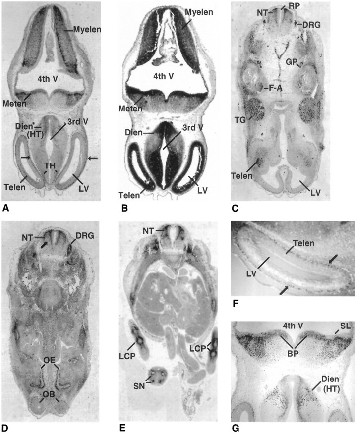
Fig. 2.

E12 expression of Olf-1 in the mouse. Low-magnification views of transverse sections (dorsal side attop) of an E12 mouse stained with the Olf-1 antibody (A, most anterior; C–E, most posterior;B, hematoxylin/eosin). The cell-dense, ventricular layer in the major brain vesicles is denoted by the strong black staining inB. Cells outside of this layer are stained a gray color (B). Olf-1 immunoreactivity in the mantle layer of the telencephalon is indicated by arrows in A andF. F and G are higher-magnification views of the section in A. Neural structures are indicated as follows: telencephalon (Telen), diencephalon (Dien), metencephalon (Meten), myelencephalon (Myelen), basal plate (BP), neural tube (NT), trigeminal ganglion (TG), facioacoustic ganglion complex (F-A), glossopharyngeal nerve (GP), dorsal root ganglia (DRG), olfactory epithelium (OE), olfactory bulb (OB), segmental nerves (SN), fourth ventricle (4th V), third ventricle (3rd V), lateral ventricle (LV), roof plate (RP), and sulcus limitans (SL). Olf-1-positive cells were also detected in the precartilage primordium of the limbs (LCP).
