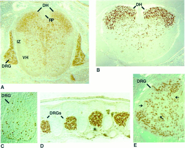
Fig. 4.
Embryonic expression of Olf-1 protein in the spinal cord and DRG. Transverse sections (dorsal side attop) of the spinal cord in an E12 (A) and PD1 (B) mouse stained with the Olf-1 antibody. Intense Olf-1 staining was detected predominantly in sensory neurons of the dorsal horns (DH), whereas only a few cells in the intermediate zone (IZ) and ventral horns (VH) were Olf-1-positive. No Olf-1 staining was observed in the roof plate (RP). Sagittal views of the dorsal root ganglia (DRG) revealed Olf-1 expression at E11 (C), E12 (A), E14 (D), and E16 (E). C and E are high-magnification views.

