
Fig. 8.
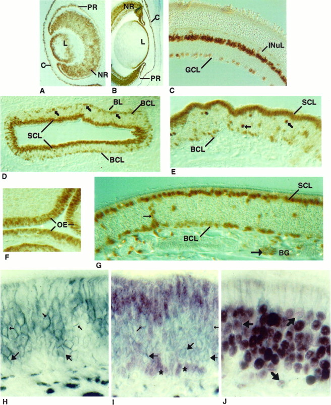
Cellular localization of Pax-6 in the retina and olfactory epithelium. Pax-6 protein was detected in the eye at E12 (A) and E16 (B) and in the adult mouse (C). The pigmented and neural retina (PRand NR), lens (L), cornea (C), ganglion cell layer (GCL), and inner nuclear layer (INuL) are indicated. In a cross-section of a PD1 mouse olfactory turbinate (D), Pax-6 immunoreactivity was observed in the sustentacular cell layer (SCL) and in the basal cell layer (BCL) (indicated by arrows). At higher magnification (E), Pax-6 immunostaining revealed additional unidentified cells in the middle of the epithelium (indicated byarrows). Pax-6 immunostaining at E12 (F) and adult (G). In G, Pax-6 protein is seen in cells of the Bowman’s glands (BG), including duct cells (small arrows) and acinar cells (large arrows). Cross-sectional views of the olfactory epithelium of a PD1 mouse immunostained with an NCAM antibody (H), Pax-6 and NCAM antibodies (I), or Olf-1 and NCAM antibodies (J). NCAM antibody stained (blue-gray) olfactory neuronal membranes (small arrows) and globose cell membranes (large arrows) inH and I. Two clusters of Pax-6 immunoreactive basal cell nuclei (red-purple) are indicated withasterisks (I). No cells were detected that reacted with both antibodies (I). Double-immunolabeled cells (J), indicated withbold arrows, were observed when the epithelium was first incubated with Olf-1 antibody (red-purple) and then with the NCAM antibody (blue-gray).