
Fig. 11.
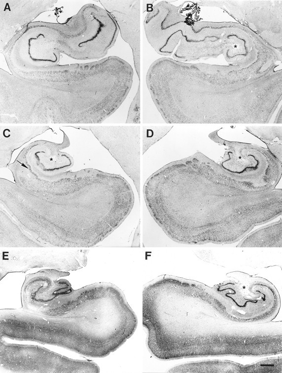
Coronal sections through the left and right sides of WH’s hippocampal formation at rostral (A, B), mid (C, D), and caudal (E, F) levels. In rostral hippocampus, more CA pyramidal cell loss is apparent on the left side (A) than on the right (B). Note the increased vascularization and the hole in the hilar region (indicated in each section on the right with a star). On the left side (A), substantial loss of neurons is apparent in the subiculum and entorhinal cortex. In a midrostrocaudal level of the hippocampus, a hole is evident in the hilar region bilaterally (stars), and increased vascularization is evident on both sides (C, D). In addition, damage is apparent in the hippocampal pyramidal cell fields and the dentate gyrus, including extensive pyramidal cell loss in the CA1 and CA3 fields, dispersion of the granule cell layer, and complete loss of polymorphic cells. Note the sparing of the superficial portion of the CA1 pyramidal cell layer on the left (arrow in C). Patchy cell loss is evident in layers III, V, and VI of the entorhinal cortex. The general pattern of damage continues through the caudal hippocampus, with increasing cell loss in the subiculum (E, F). Some preservation of CA1 and CA2 pyramidal cells is visible on the right side (F). Scale bar, 1 mm.