Fig. 1.
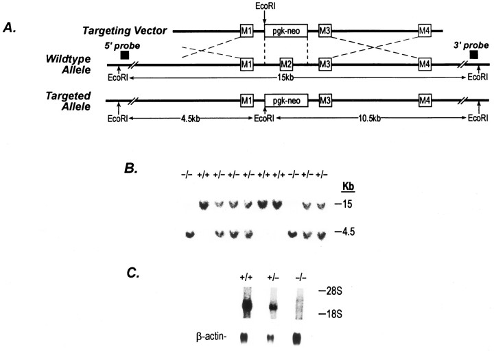
Targeted disruption of the NR2C gene.A, Schematic representations of the targeting vector, wild-type allele, mutant allele, and the strategy for NR2C knockout. Four putative segments (M1–M4) are shown as open boxes. The location of the 5′- and 3′-external probes is shown. Both probes hybridize to a 15 kb EcoRI fragment from the wild-type allele and to 4.5 and 10.5 kb EcoRI fragments from the mutated allele, respectively. B, Southern blot analysis of progeny from intercross of heterozygotes (NMDA 2C +/−). 5′-External probe was used in this experiment. A similar result was obtained using the 3′-external probe (data not shown). C, Northern blot analysis of wild-type and mutated mice. Poly(A+) RNA was isolated from total brain tissue from 4-d-old animals. RNA was separated in a formaldehyde gel and transferred to a nylon membrane (Hybond-N+, Amersham). Membrane was hybridized with a fragment of NR2C cDNA containing all four transmembrane segments. The same filter was rehybridized with a β-actin probe.

