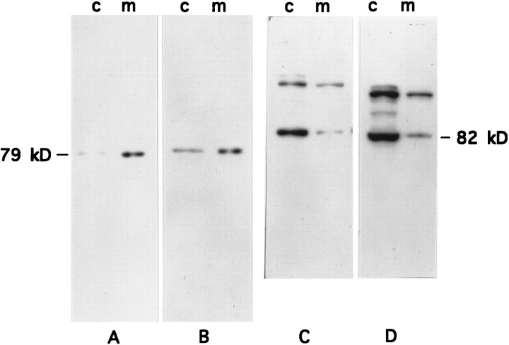
Fig. 2.
Subcellular distribution of PKC θ and α isoforms in primary rat myotubes. Primary myoblast cultures were established and cultured for 8 d to permit differentiation into myotubes. Myotube cultures were homogenized and separated into cytosolic (c) and membrane (m) protein fractions by ultracentrifugation. Fractions were adjusted either to equal protein concentration (A, C, 100 μg/lane) or to equal volume (B, D, 100 μl/lane) and were separated by SDS-PAGE and transferred to nitrocellulose membranes. The immunoblots were probed with anti-nPKC θ antiserum (A,B) or anti-cPKC α antibody (C, D) as described in the legend to Figure 1 and were exposed for autoradiography. nPKC θ and cPKC α migrate as 79 and 82 kDa proteins, respectively.

