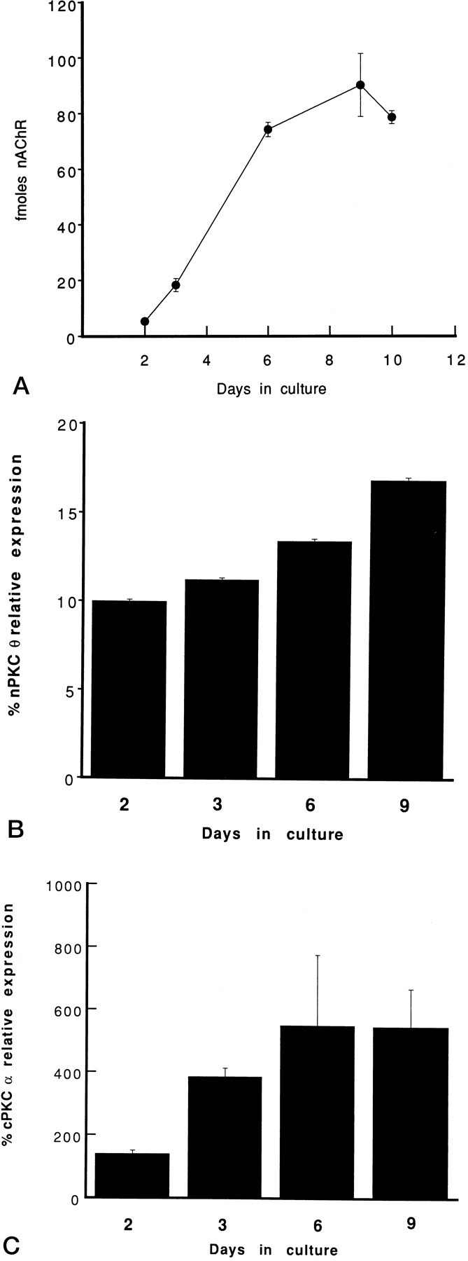
Fig. 3.

Time course of the expression of nAChRs and PKC α and θ isoforms during myogenic differentiation in culture. Primary myoblast cultures were established in parallel. At the indicated days in culture, cells were solubilized and specific binding of [125I]α-bungarotoxin to nAChRs was quantitated. Data are expressed as fmol of nAChR per 35 mm tissue culture dish (A). Alternatively, cells were homogenized and membrane (B) or cytosolic (C) proteins were separated by SDS-PAGE and transferred to nitrocellulose membranes. The immunoblots were probed with anti-PKC θ antiserum (B) or anti-PKC α antibody (C) and analyzed according to the legend to Figure 1. PKC isoform levels in membrane or cytosolic subcellular fractions (100 μg/lane) of cultured cells are expressed as percent PKC isoform expression in membrane (B) or cytosolic (C) protein (100 μg/lane) fractions of adult skeletal muscle, respectively. Each experiment was performed in triplicate. Error bars represent SEM.
