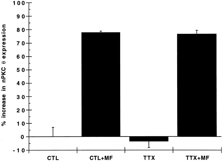
Fig. 9.
Effect of tetrodotoxin on myotube nPKC θ expression. Primary myoblast cultures were established and cultured for 7 d as described in the legend to Figure 2. Myotubes were preincubated for 3 hr with (TTX) or without (CTL) 500 nm tetrodotoxin. The subcellular membrane fraction from 1 × 10⁶NG108-15 cells (+MF) or buffer alone was added, and the cultures were incubated for 2 d in the continued presence of 500 nm tetrodotoxin. Myotube cultures were homogenized, and equal protein (100 μg) was analyzed as described in the legend to Figure 1. Data are expressed as percent increase in nPKC θ expression in cytosolic plus membrane fractions of experimental compared with control (CTL) cultures. Each experiment was performed in triplicate. Error bars represent SEM.

