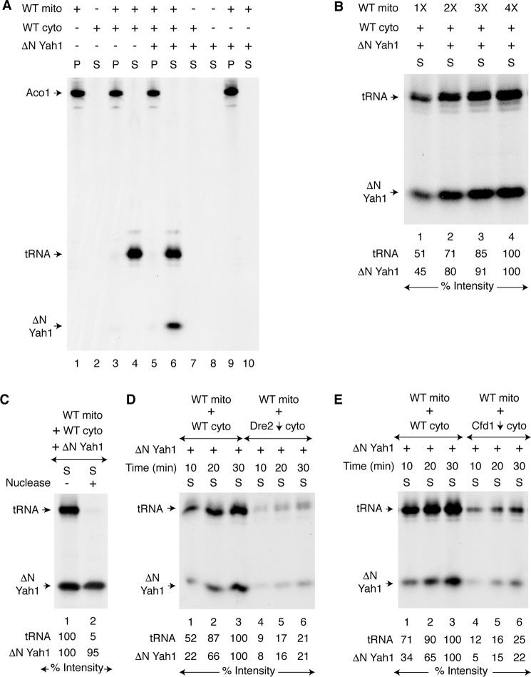
Figure 1.
Requirement of mitochondria for cytoplasmic iron–sulfur cluster assembly. A, WT mitochondria (200 μg of proteins) alone, WT cytoplasm (200 μg of proteins) alone, or both were mixed with [35S]cysteine (10 μCi), nucleotides (1 mm GTP, 2 mm NADH, and 4 mm ATP), iron (10 μm ferrous ascorbate), and as indicated, apo-ΔN60 Yah1 protein (1 μg). The samples were incubated at 30 °C for 30 min. After centrifugation, the pellet (P; mitochondria) and supernatant (S; cytoplasm) fractions were analyzed by native PAGE followed by autoradiography. B, WT mitochondria (1× = 100 μg of proteins) were supplemented with WT cytoplasm (200 μg of proteins), apo-ΔN60 Yah1, [35S]cysteine, nucleotides, and iron. After incubation at 30 °C for 30 min, the samples were centrifuged, and the cytoplasm/supernatant (S) fractions were analyzed. C, reaction mixtures containing WT mitochondria, WT cytoplasm, apo-ΔN60 Yah1, [35S]cysteine, nucleotides, and iron were incubated at 30 °C for 30 min. After centrifugation, the cytoplasm/supernatant (S) fraction was treated with S7 micrococcal nuclease (800 units/ml) at 30 °C for 10 min as indicated and analyzed. D, WT mitochondria were added to WT cytoplasm or Dre2-depleted (Dre2↓) cytoplasm. The samples were incubated with apo-ΔN60 Yah1, [35S]cysteine, nucleotides, and iron at 30 °C for 10–30 min as indicated. After centrifugation, the cytoplasm/supernatant (S) fractions were analyzed. E, WT mitochondria were added to WT cytoplasm or Cfd1-depleted (Cfd1↓) cytoplasm, and assays were performed as in D. WT mito, WT mitochondria; WT cyto, WT cytoplasm.

