Figure 3.
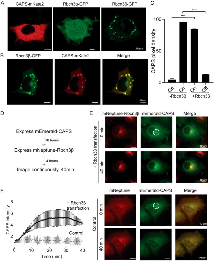
Rbcn3β expression in COS cells recruits CAPS1 to membrane structures. A, representative epifluorescence images of COS cells expressing CAPS1-mKate2, Rbcn3α-GFP, or Rbcn3β-GFP. B, representative confocal image of COS cells coexpressing Rbcn3β-GFP and CAPS1-mKate2 showing that CAPS1-mKate2 is recruited to Rbcn3β-containing membranes. C, studies were quantified by determining mean pixel intensity of CAPS1-mKate2 on Rbcn3β-GFP+ structures (On) or in cytosol (Off). Values represent means ± S.E. of triplicate cells in five independent experiments (****, p < 0.00005). D, sequential transfection scheme for investigating the recruitment of mEmerald-CAPS to mNeptune-Rbcn3β membrane structures. E, upper panels, epifluorescence images of mEmerald-CAPS–expressing COS cell (middle panels) after 4 h of mNeptune-Rbcn3β expression (upper panels) and 40 min later (lower panels). Lower panels, similar study to that shown in upper panels but with mNeptune expression. F, to quantify the ongoing recruitment of mEmerald-CAPS to mNeptune-Rbcn3β membranes, fluorescence was quantified within the indicated region of interest in E over 40 min for mNeptune-Rbcn3β– and mNeptune-expressing cells with 0-min background values subtracted. Values shown represent means ± S.E. of three independent studies (n = 3).

