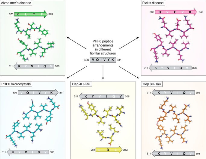
Figure 4.
Different arrangements of the PHF6 peptide in the different fibrillar structures. The peptide is indicated as a gray arrow, with inward-pointing residues in white and outward-pointing residues in black. Structures are from the AD PHFs (top left, green), the PiD PHFs (top right, magenta), the PHF6 microcrystals (bottom left, blue), the heparin-induced 4R-Tau structure (bottom middle, yellow), and the heparin-induced 3R-Tau fibers (bottom right, orange). The peptide itself adopts invariably the same extended conformation but faces different peptides in every single structure. In the heparin-induced 4R-Tau fibrils, Lys311 points in the same direction as Tyr310, and both residues face the outside of the fibrillar structure.

