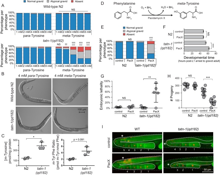
Figure 4.
tatn-1 mutants are sensitive to the adverse effects of m-tyrosine. A, treatment of N2 and tatn-1(qd182) mutant worms with varying concentrations of either p- or m-tyrosine revealed a decrease in fertility of the tatn-1 mutants supplemented with m-tyrosine. Approximately 150 worms were scored per treatment, and the percentage of worms showing each reproductive phenotype—based on the number of eggs in the uterus of day 1 adult animals with normal gravid showing ≥6 embryos within the uterus, abnormal gravid showing <6 embryos, and absent showing zero embryos—are graphed. ***, p < 0.001 for χ2 test of the effects of equal concentrations of m- and p-tyrosine within the same strain. ††, p < 0.01; †††, p < 0.001 for χ2 test of the effects of m-tyrosine treatment of equal concentrations between strains. B, representative images of worms quantified in A. C, results of LC/MS-MRM quantification of the concentration of free m-tyrosine and the m-tyrosine/phenylalanine ratio for N2 and tatn-1(qd182) mutant worms grown on NGA plates supplemented with 4 mm m-tyrosine. Approximately 1500 worms were collected per biological replicate, and the amino acid concentrations were normalized to the concentration of soluble worm protein measured per sample. *, p < 0.05 by t test. D, diagram of the enzymatic formation of m-tyrosine from phenylalanine by the pacidamycin X enzyme produced by Streptomyces coeruleoribudus. E, treatment of N2 and tatn-1(qd182) mutant worms fed bacteria expressing PacX or an empty vector control revealed a decrease in fertility in the tatn-1 mutants treated with PacX-expressing bacteria. Scoring and statistical analysis were performed as in A. F, results of a developmental time assay in which the time required for L1 arrested N2 and tatn-1(qd182) animals treated with control or PacX-expressing bacteria to become gravid adults is measured. 4–5 worms were assayed per condition. Mean developmental time and S.D. (error bars) are shown. ***, p < 0.001; NS, nonsignificant p value by t test. G, measurement of the percentage of embryonic lethality in the worms from F treated with either control or PacX-expressing bacteria demonstrates an increase only in the tatn-1 mutants treated with the PacX-expressing bacteria. Shown are the mean percentage of embryonic lethality from each animal and the S.D. ***, p < 0.001; NS, nonsignificant p value by t test. H, measurement of the fertility of N2 and tatn-1(qd182) worms treated with control or PacX-expressing bacteria. n = 9–10 worms assayed per genotype and treatment. Shown are the mean number of progeny and S.D. for each genotype-condition pair. ***, p < 0.001; NS, nonsignificant p value by t test. I, representative images of WT and tatn-1(qd182) transgenic worms expressing fluorescent proteins that label the cell membrane (green) and nucleus (red) of the developing oocytes. The germline is outlined with a solid line, the worm body is outlined with a dashed line, mature oocytes are numbered, and a star marks the beginning of oocyte expansion. The WT worms exhibit normal gonadal architecture regardless of treatment. The control-treated tatn-1 mutant worm shows a delay in oocyte expansion. The PacX-treated tatn-1 mutant also shows a delay in oocyte expansion along with a reduction in mature oocytes and a defect where the germline fails to cross the midline.

