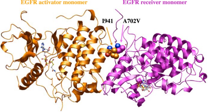
Figure 6.

Effect of EGFR A702V on kinase interface. Modeled A702V (receiver kinase) interaction with Ile-941 (activator kinase) at the asymmetric dimer interface in the 2.6 Å resolution EGFR X-ray structure with bound ATP peptide analogue (PDB code 2GS6). A structural water (small red sphere) is nearby. Carbon atoms for the Ile-941 and A702V are shown as orange and magenta spheres, respectively; the red and blue spheres, where seen, correspond to main-chain carbonyl oxygen and amino groups, respectively.
