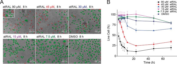
Figure 1.
Image-based evaluation of nuclear morphology in U2OS cells after atRAL exposure. A, apoptotic changes in nuclear morphology observed by Hoechst 33342 staining. Shown are representative images of the nuclear morphology counterstained with Hoechst 33342 after an 8-h incubation with atRAL at 7.5, 15, 30, 45, and 90 μm or with DMSO. Nuclear morphology was assessed with Harmony® high-content imaging and analysis software. Red, dead cells, nuclear area < 200 pixels2; green, live cells, nuclear area > 200 pixels2. B, nuclear morphology quantification monitored through 3 days post-atRAL at 7.5, 15, 30, 45, and 90 μm or with DMSO. Error bars, S.D. of triplicate readings.

