. 2019 May 22;8:e44904. doi: 10.7554/eLife.44904

Figure 3. Nmd3 interacts with Lsg1 and eIF6 throughout 60S maturation.

(A) Conformation of H69 (states I-VI) differs from the mature 60S subunit (4v88, Ben-Shem et al., 2011). (B) Interaction of Lsg1 with the tip of H69. (C) Contact between Nmd3 residue N281 and base U2269 (H69). (D) Interactions of Nmd3 SH3 domain with H68 and H74. (E) Interaction between Lsg1 and the Nmd3 eL31-like domain. (F) Interaction of the Nmd3 N-terminal domain with the D and E domains of eIF6.

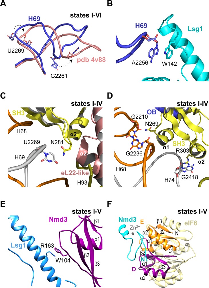
Figure 3.

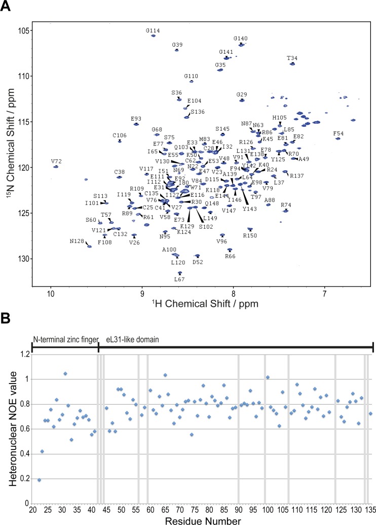
Figure 3—figure supplement 1. NMR spectroscopic analysis of A.fulgidus Nmd3.

Figure 3—figure supplement 1.

(A) Assigned 1H-15N BEST-TROSY spectrum of residues 22–150 in solution. (B) Heteronuclear 1H{15N} NOE analysis of A. fulgidus Nmd3. Values close to one indicate rigid secondary structure and values < 1 have individual flexibility on the picosecond timescale. Exchange broadened residues are marked with grey bars.