. 2019 May 22;8:e44904. doi: 10.7554/eLife.44904

Figure 6. Mechanism of Nmd3 release.

(A) The Nmd3 β4–5 loop masks base A2971 (state IV). Insert shows base A2971 in the ‘closed’ position. H38 is ‘open’, the L1 stalk is ‘closed’ and uL16 is loaded. The A, P and E tRNA binding sites are indicated. (B) The uL16 P-site loop competes with the Nmd3 β4–5 loop for binding to base A2971 (state V). The L1 stalk is partially retracted (rotated by 20° relative to the fully closed L1 stalk); the Nmd3 β4–5 loop is displaced from the PTC. The uL16 P-site loop is bound to base A2971 in the flipped-out conformation. (C) The completed PTC (state VI). The L1 stalk is fully open (rotated by 56°), Nmd3 and Lsg1 have dissociated and the uL16 P-site loop is bound to base A2971 in the flipped-out conformation. (D) L1 stalk retraction promotes a conformational switch in the Nmd3 eL22-like domain. Atomic models for the L1 stalk and Nmd3 in states IV (magenta) and V (gold) are superimposed. The rotational pivot point between the eL31- and eL22-like domains is indicated.

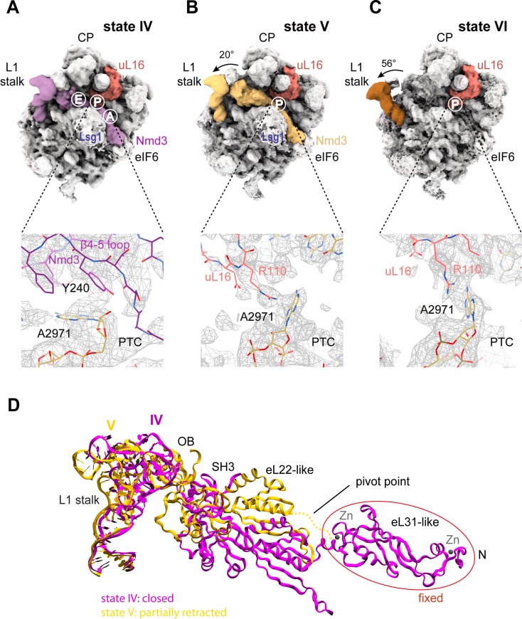
Figure 6.

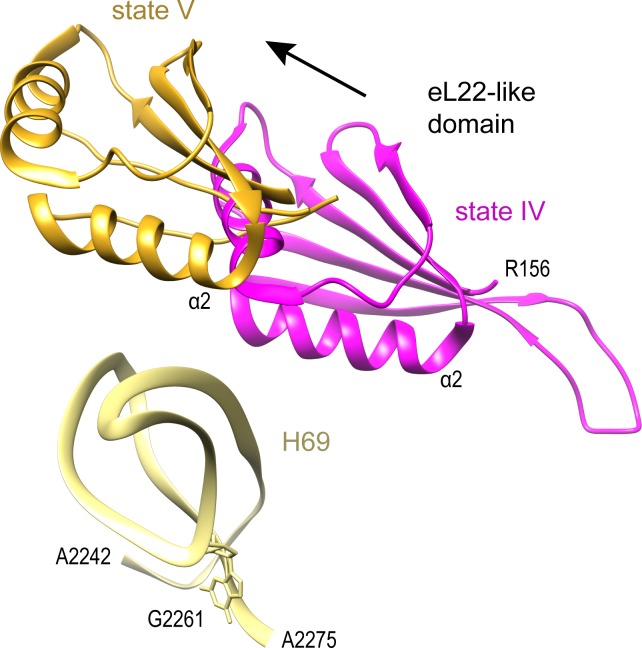
Figure 6—figure supplement 1. Release of Nmd3 eL22-like domain involves new contacts with H69.

Figure 6—figure supplement 1.

Nmd3 eL22-like domain is shown in magenta (state IV) and gold (state V), H69 is yellow.
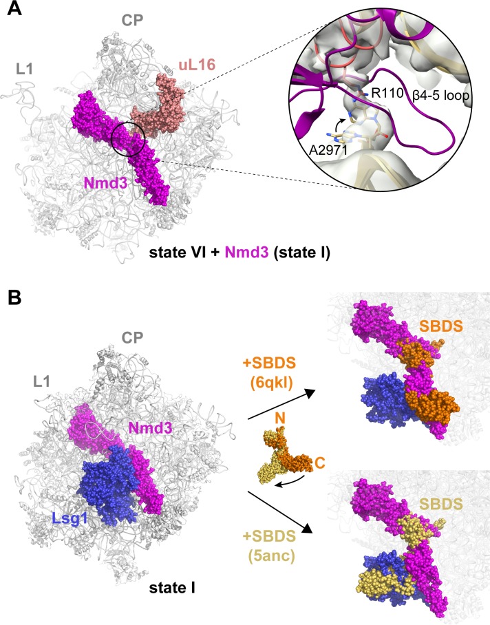
Figure 6—figure supplement 2. Structural conflicts during the completion of PTC assembly.

Figure 6—figure supplement 2.

(A) Structural conflict between the Nmd3 β4–5 loop and the uL16 P-site loop. Inset shows the Nmd3 β4–5 loop (magenta) from state IV superimposed with the uL16 P-site loop (dark salmon) from state V bound to base A2971 in the flipped-out conformation. Arrow indicates the change in conformation of base A2971 between states IV-V. (B) Lsg1 and Nmd3 are placeholders for SBDS. SBDS (pdb 6qkl, 5anc) is superimposed with the cryo-EM density map of state I. Cryo-EM density for Lsg1 is blue, Nmd3 is magenta, SBDS (pdb 6qkl) is brown, SBDS (pdb 5anc) is gold.
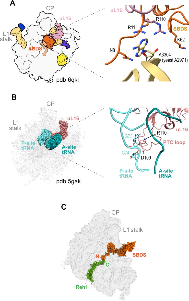
Figure 6—figure supplement 3. Quality control assessment of PTC assembly by SBDS.

Figure 6—figure supplement 3.

(A) Interactions between uL16 (dark salmon), SBDS (light orange) and Dictyostelium base A3304 (equivalent to yeast A2971) (gold). (B) Interactions of uL16 with the A site (dark green) and P site (cyan) tRNAs (pdb 5gak). (C) Potential cross-talk between SBDS and Reh1 in the peptide exit tunnel. The cryo-EM density for SBDS (orange, pdb code 6qkl) is superimposed with the cryo-EM density map of state VI. Reh1 density is green.
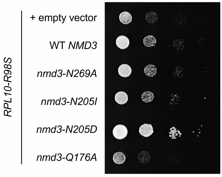
Figure 6—figure supplement 4. Suppression of the T-ALL associated uL16-R98S variant by mutations in NMD3.

Figure 6—figure supplement 4.

Complementation of GAL10::RPL10 rpl10-R98S cells with nmd3 variant alleles. Ten-fold serial dilutions (from left to right) of the indicated strains are shown.