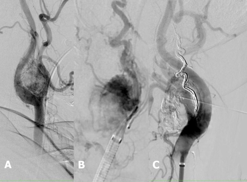
Figure 2. (A) Pre-embolization angiography showing the hypervascularized tumor and a left carotid lyre sign; (B) Superselective catheterization of the glomus tumor and “pressure cooker” embolization; (C) Direct external puncture of the glomus tumor and embolization.