Figure 1.
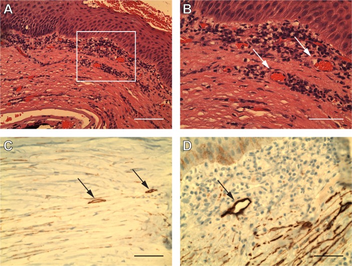
Neovascularisation and suspected lymphatics within failed corneal grafts. (A, B and D) Case ID #1: an 88 year-old male patient with failed graft. (C) Case ID #2: an 87-year-old female patient with failed graft. All nine cases of corneal graft failure were found to contain neovascularisation, which is demonstrated above on H&E (A and B). Blood vessels are outlined by a rectangular area shown at 20× magnification (A). Blood vessels are indicated by arrows at 40× magnification (B). Monocellular infiltrates were seen around the lumen of these vessels. All nine cases of corneal graft failure were found to contain luminal and elongated profiles via podoplanin antibody (D2-40) immunohistochemistry. Two immunoperoxidase images are provided above with podoplanin-antibody staining lymphatics denoted by arrows (C and D). Scale bars represent (A) 100 µm and (B–D) 50 µm.

