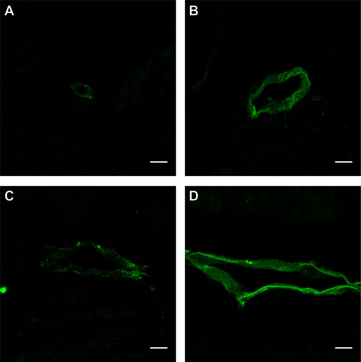
Figure 4.
Lymphatics of varying morphologies and sizes within a single case. Case ID # 1: the above immunofluorescence images were obtained from an 88-year-old male patient with a failed graft. Distinct podoplanin-positive lymphatics with different sizes, morphologies and clear lumen from one single case are shown (A–D) at 63× magnification. The largest measured diameters of the lymphatics shown were as follows: (A) 9 µm, (B) 33 µm, (C) 50 µm, (D) 84 µm. All scale bars for this figure represent 10 µm.

