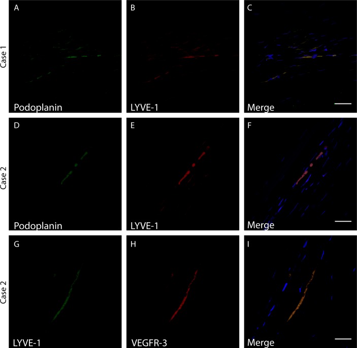
Figure 6.
Fluorescence in situ hybridisation detection of lymphatic marker mRNAs. (A–C) Case ID #2: an 87-year-old female patient with a failed graft. (D–I) Case ID #5: a 30-year-old male patient with a failed graft. Probes recognising (A and D) podoplanin mRNA, (B, E, and G) LYVE-1 or (H) VEGFR-3 were used. (C, F and I) 4′,6-diamidino-2-phenylindole is also shown (blue). A merged image (C, F) illustrates podoplanin and LYVE-1 double positive regions (yellow). Another merged image (I) illustrates a LYVE-1 and VEGFR-3 double positive region (orange). Scale bars represent 30 µm.

