
Figure 3. Adipocyte derived exosomes transport neutral lipid.
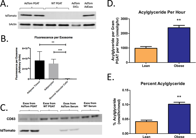
(A) Western blot of total protein from whole AdTom PGAT, WT PGAT, SVCs isolated from AdTom PGAT, and AdExos isolated from AdTom PGAT. Blots were probed using antibodies against tdTomato and bActin. (B) TdTomato fluorescence per exosome, as measured by Nanoparticle Tracking Analysis, for AdExos purified from whole AdTom PGAT, adipocytes isolated from AdTom PGAT, and SVCs isolated from AdTom PGAT (One-way ANOVA. n = 4, ** p-value < 0.01, *** p-value < 0.001). (C) Western blot of total protein from AdExos isolated from AdTom PGAT, AdExos isolated from WT PGAT, exosomes isolated from AdTom serum, and exosomes isolated from WT serum. Blots were probed using antibodies against CD63 and tdTomato. (D) Acylglyceride content of purified adipocyte-derived exosomes from lean and obese (Lepob/ob) adipose tissue per gram of PGAT per hour (Unpaired two-tailed t-test. n = 8; ** p-value < 0.01). (E) Acylglyceride content of purified adipocyte-derived exosomes, expressed as a percentage of the acylglyceride content of the tissue itself (Unpaired two-tailed t-test. n = 8; ** p-value < 0.01).