Figure 6.
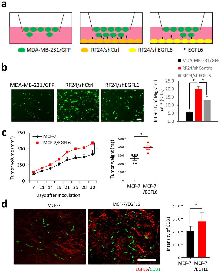
EGFL6 expression in endothelial cells promotes breast cancer cell migration and increases tumor angiogenesis. a, Diagram shows the layout of co-culture assays. b, Endothelial cells (RF24) promoted migration of cancer cells. MDA-MB-231 cancer cells were seeded on transwell upper inserts and RF24/shControl and RF24/shEGFL6 cells were seeded in the lower chamber. After 8 hours incubation, migrated cancer cells were imaged and quantified in the bar graph. A representative image for each conditions is shown from 3 replication, n=3. Scale bar, 50 μm c, MCF-7/EGFL6 cells showed increased tumor growth than the parental control. Tumor sizes were measured in the indicated days. Tumor weights were measured at the end point of the experiment. d, EGFL6 expression in cancer cells promoted tumor angiogenesis in vivo. IF staining of EGFL6 (in red) and CD31 (green) in tumor tissues, representative images with overlay of nuclear staining (blue) in MCF7/EGFL6 and MCF7 xenograft tumor tissues are shown and quantified. Scale bar, 100 μm. All experiments were repeated 3 to 5 times, *, p < 0.05. Error bar, SD.

