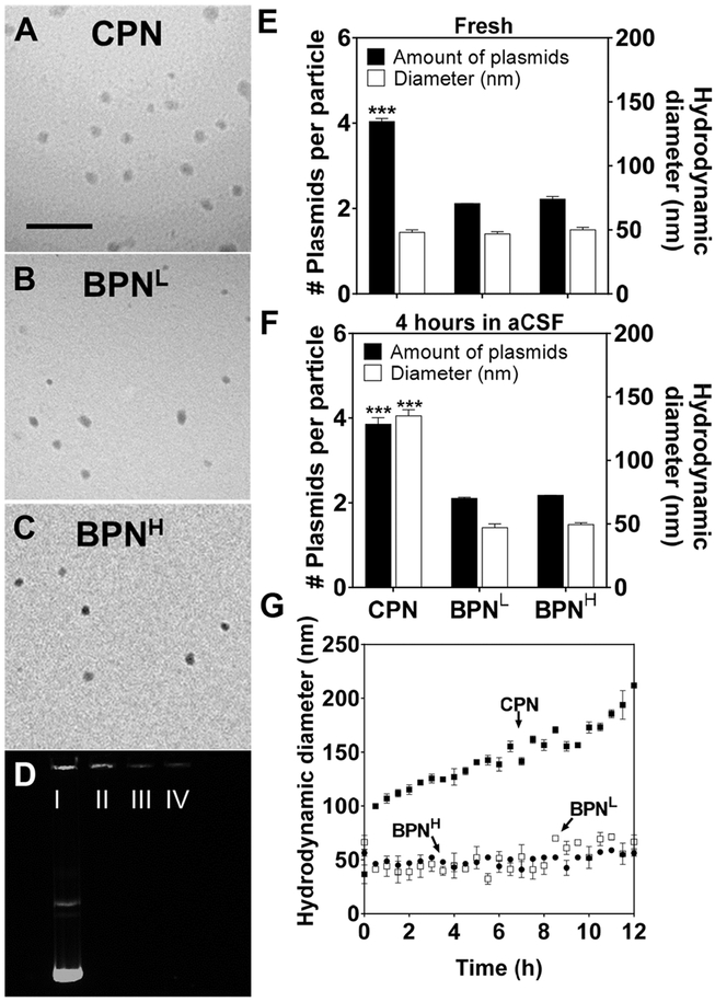
Figure 1. Physicochemical properties and stability of DNA-NP.
Transmission electron microscopy images of (A) CPN, (B) BPNL, (C) BPNH freshly made in ultrapure water. Scale bar = 400 nm. and (D) Compaction of plasmids in DNA-NP shown by electrophoresis: I) free DNA, II) CPN, III) BPNL, IV) BPNH. Number of plasmids in each DNA-NP and hydrodynamic diameter of DNA-NP when (E) freshly made in water and (F) when incubated in aCSF for 4 hours. (G) Change in hydrodynamic diameters over 12 hours in aCSF at 37°C measured by DLS. Differences are statistically significant as indicated (***p < 0.001), of CPN compared to BPNL and BPNH.

